Loculated Pleural Effusion X Ray : The management of benign non-infective pleural effusions ... / A pleural effusion is an abnormal collection of fluid within the pleural space.
byAdmin-
0
Loculated Pleural Effusion X Ray : The management of benign non-infective pleural effusions ... / A pleural effusion is an abnormal collection of fluid within the pleural space.. If you miss a tension pneumothorax you risk your patient's. He has a vasculitic peripheral rash and feels generally unwell. This position is called lateral decubitus position. Pleural effusions can loculate as a result of adhesions. Pleura is a mesothelial lined sac that envelopes the lungs and comprises of 2 membranous walls i.e.
In the usa approximately 1.5 million people are diagnosed with a pleural effusion each year 2. Case contributed by dr prashant mudgal. Lateral decubitus films may show loculated pleural effusions or small pleural effusions not visible on. 304 187 просмотров 304 тыс. The patient's history and physical exam may indicate a presumptive.
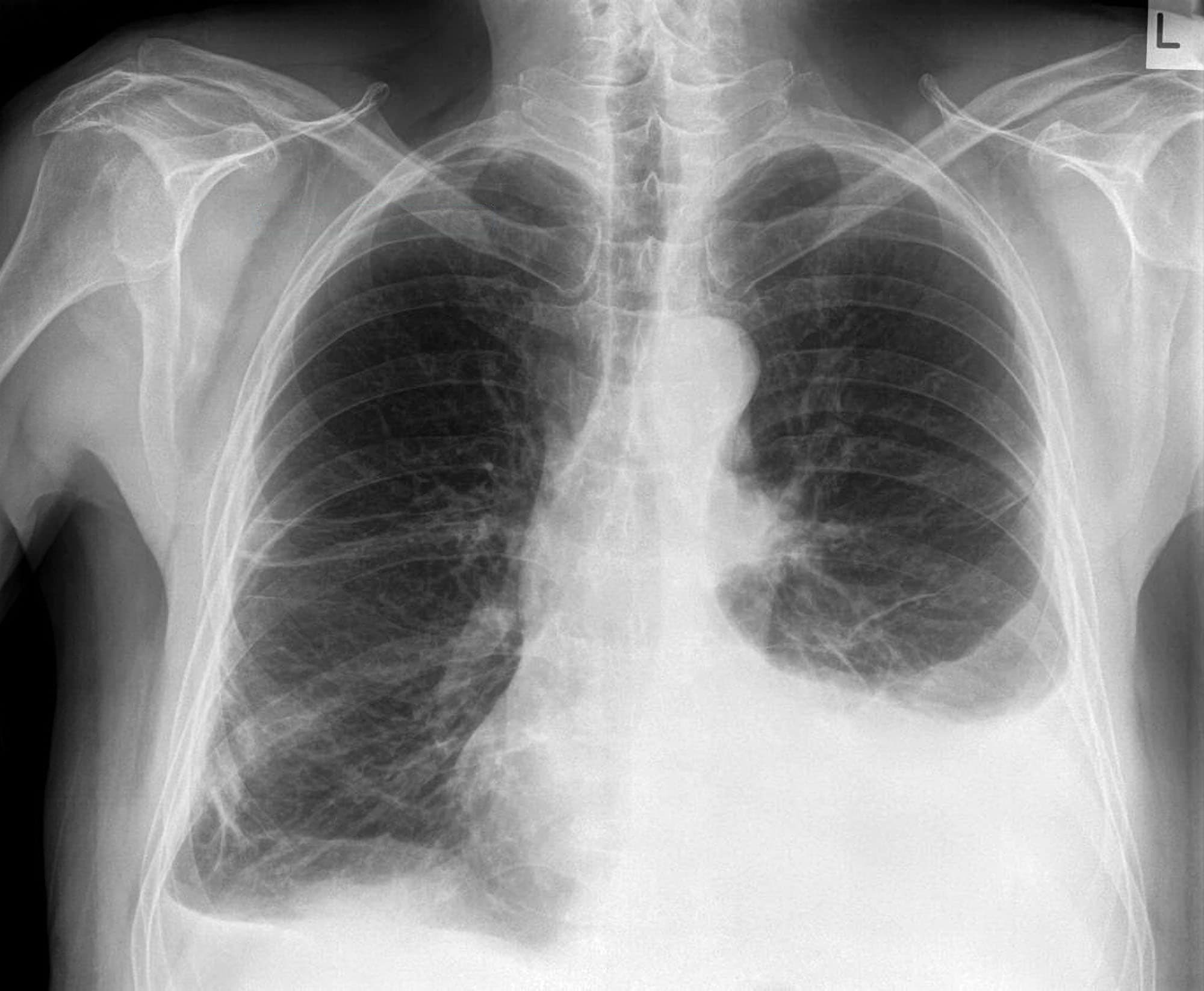
Pleural Effusion | Chest X-Ray - MedSchool from medschool.co Obliteration of left costophrenic angle with a wide pleural based dome shaped opacity projecting into the lung noted tracking along the cp angle and lateral. The second effusion is loculated. In the usa approximately 1.5 million people are diagnosed with a pleural effusion each year 2. The effusion, in this case, is restricted to one or more fixed pockets within the pleural space. A pleural effusion is accumulation of excessive fluid in the pleural space, the potential space that surrounds each lung. Although pleural effusion volumes can be estimated by visual inspection with good correlation, some overestimation is consistently seen. Us scan they can be identified clearly and it is very complicated.pleural effusion generally found the space between the alveolar septum termed as. Excluding the loculated effusions, the coefficient of correlation was 0.969 for the right side and 0.949 for the left side (p<.001).
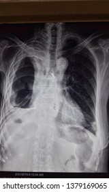
The plain chest radiographic features of pleural effusion are usually characteristic.
It allows distinction between free and loculated fluid showing its extent and localization. A role in selected clinical circumstances. Pleural effusions can loculate as a result of adhesions. Treatment of loculated pleural effusions with. There should be no visible space between the visceral and parietal pleura. Pleural effusions accompany a wide variety of disorders of the lung, pleura, and systemic disorders. He has a vasculitic peripheral rash and feels generally unwell. Pleura is a mesothelial lined sac that envelopes the lungs and comprises of 2 membranous walls i.e. Pleural fluid studies were suggestive of a transudative process, though with some abnormal characteristics (including lymphocyte predominance, as well as presence of signet cells). A pleural effusion is an abnormal collection of fluid within the pleural space. The patient's history and physical exam may indicate a presumptive. Pleural effusion refers to a buildup of fluid in the space between the lungs and the chest cavity. Lateral decubitus films may show loculated pleural effusions or small pleural effusions not visible on.
Pleural effusions can loculate as a result of adhesions. Ct scan is the most sensitive modality for detection of presence of minimal fluid. 304 187 просмотров 304 тыс. Suspected parenchymal or pleural pathology. This position is called lateral decubitus position.
Loculated Pleural Effusion Images, Stock Photos & Vectors ... from image.shutterstock.com If you miss a tension pneumothorax you risk your patient's. Concave meniscus (horizontal in case of. The patient's history and physical exam may indicate a presumptive. What procedures and tests diagnose pleural effusions? A parasternal long axis and subcostal views are shown. Treatment of loculated pleural effusions with. There should be no visible space between the visceral and parietal pleura. Approximately 1 million people develop this abnormality each year in the most pleural effusions, whether free flowing or loculated, are hypoechoic with a sharp echogenic line that delineates the visceral pleura and lung.
Ct scan is the most sensitive modality for detection of presence of minimal fluid.
The annual incidence of pleural effusion in the developed world has been estimated at 320 per 100,000 population per year 1. The pleura and pleural spaces are only visible when abnormal. A parasternal long axis and subcostal views are shown. Ct scans show more detail than. Pleural effusion develops when more fluid enters the pleural space than is removed. Us scan they can be identified clearly and it is very complicated.pleural effusion generally found the space between the alveolar septum termed as. A pleural effusion is accumulation of excessive fluid in the pleural space, the potential space that surrounds each lung. The second effusion is loculated. The patient's history and physical exam may indicate a presumptive. Suspected parenchymal or pleural pathology. Concave meniscus (horizontal in case of. Pleural effusions accompany a wide variety of disorders of the lung, pleura, and systemic disorders. Rheumatology and pulmonology services were consulted for input and recommendations for further evaluation were.
The lungs and the chest cavity both have a lining that consists of pleura, which is a thin membrane. The plain chest radiographic features of pleural effusion are usually characteristic. Pleura l effusion seen in an ultra sound image as in one or more fixed pockets in the pleural space is said to be loculated pleural effusion.in. A pleural effusion is accumulation of excessive fluid in the pleural space, the potential space that surrounds each lung. Ct scan is the most sensitive modality for detection of presence of minimal fluid.
Loculated pneumothorax | Radiology Case | Radiopaedia.org from images.radiopaedia.org Pleura is a mesothelial lined sac that envelopes the lungs and comprises of 2 membranous walls i.e. Concave meniscus (horizontal in case of. Suspected parenchymal or pleural pathology. Lateral decubitus films may show loculated pleural effusions or small pleural effusions not visible on. He has a vasculitic peripheral rash and feels generally unwell. Surgical thoracostomy tube placement and radiologically guided catheter drainage are standard therapy for loculated pleural fluid loculated pleural effusion radiology case radiopaedia.org. A parasternal long axis and subcostal views are shown. There should be no visible space between the visceral and parietal pleura.
Although pleural effusion volumes can be estimated by visual inspection with good correlation, some overestimation is consistently seen.
Pleural fluid studies were suggestive of a transudative process, though with some abnormal characteristics (including lymphocyte predominance, as well as presence of signet cells). Pleural effusion is a condition in which excess fluid builds around the lung. What procedures and tests diagnose pleural effusions? A role in selected clinical circumstances. In healthy lungs, these membranes ensure that a. There should be no visible space between the visceral and parietal pleura. Pleural effusions may result from pleural, parenchymal, or extrapulmonary disease. Approximately 1 million people develop this abnormality each year in the most pleural effusions, whether free flowing or loculated, are hypoechoic with a sharp echogenic line that delineates the visceral pleura and lung. 304 187 просмотров 304 тыс. The annual incidence of pleural effusion in the developed world has been estimated at 320 per 100,000 population per year 1. The plain chest radiographic features of pleural effusion are usually characteristic. Although pleural effusion volumes can be estimated by visual inspection with good correlation, some overestimation is consistently seen. Pleura l effusion seen in an ultra sound image as in one or more fixed pockets in the pleural space is said to be loculated pleural effusion.in.
The pleural fluid may loculate between the visceral and parietal pleura (when there is partial fusion of the pleural layers) or within loculated pleural effusion. Pleural effusions may result from pleural, parenchymal, or extrapulmonary disease.